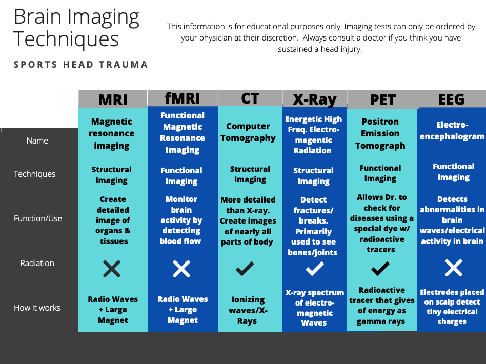
When should you get brain imaging done?
Always consult a physician if you think you have sustained a head injury. They will order imaging tests at their discretion.
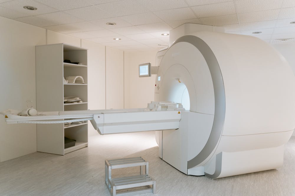
MRI- Magnetic Resonance Imaging
A physician may recommend an MRI if they suspect that a patient has a brain aneurysm, blocked arteries or a brain tumor. The 3D imaging helps detect forms of TBI and also abnormalities in the brain that could later develop into diseases like Alzheimers. An MRI creates more detailed images of organs, torn ligaments, herniated disks, and soft tissue damage compared to CT scans. They also catch inflammation better than X-rays and CT scans. This procedure is painless, and lasts between 15-90 minutes depending on how large the area being scanned is as well as the number of photos being taken.
fMRI-Functional Magnetic Resonance Imaging
An fMRI scan measures brain activity by detecting changes associated with oxygen and blood flow. Cerebral blood flow and neuron activation work in parallel which means that when a part of the brain is active, it consumes more oxygen. In order for more oxygen to get to this part of the brain, blood flow needs to be increased. The fMRI scan is used often to evaluate how well different parts of the brain are handling critical functions.
CT Scan- Computerized Tomography
A CT scan uses multiple x-ray images and computer processing to create images of bones, joints, blood vessels, and soft tissues. CT scan cross-sectional images can show complex fractures and tumors as well as internal bleeding. Head CT scans take pictures of a patient’s skull, blood vessels, brain, and sinuses. Doctors can receive test results almost immediately and are more detailed than X-ray images.
X-Ray
X-rays are not used very often for head injuries. They are able to detect skull fractures, but doctors are unable to assess injuries to the brain via this method. In this situation, a CT scan would most likely be used instead of an x-ray.
PET- Positron Emission Tomography
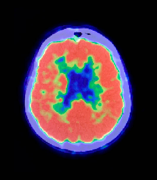
PET scans can be used to detect early signs of brain disorders, heart disease, and certain cancers. A type of dye that has radioactive tracers is either swallowed, injected, or inhaled (this depends on what part of the body the doctor is looking at). A PET scan of the brain can show your physician how your brain and tissues are functioning.
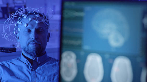
EEG-Electroencephalogram
Doctors often order an EEG test to identify causes of behavioral changes, and monitor and diagnose seizure and sleep disorders. An EEG detects abnormalities in brain waves and electrical activity measured by several electrodes placed on a patient’s scalp.